| Spondylolisthesis
shown to require additional fusion segment once its degree of instability,
not visible by recumbent-only MRI, was demonstrated by Fonar Upright
MRI.
Clinical Case Overview
The patient was a 49-year-old male who had had a 20-year history
of chronic back pain and a three-year history of right lower extremity
radiculopathy.
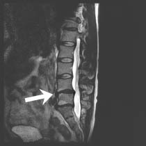
Prior to the Upright™ scan, the patient was scanned in a
recumbent-only MRI (1.5T). It showed a right paracentral disk herniation
at L5-S1. Based on the recumbent images, neurosurgeon Bennie W.
Chiles III, M.D., said:
 |
 |
 |
Neutral-Sit |
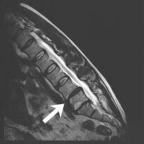
Flexion |
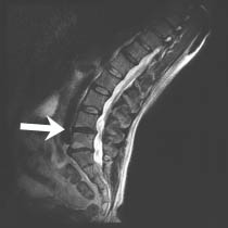
Extension |
“I would have likely performed a diskectomy at L5-S1 to relieve
pressure on the nerve root, along with an L5-S1 fusion for the back
pain. Fusing L4-5 was not an initial consideration because no spinal
instability was seen on the recumbent MRI.
When the dynamic flexion and extension images performed
in the Upright™ MRI demonstrated an instability at L4-5 and
showed the full extent of that instability once the patient’s
body weight was applied, I chose to also fuse L4-5 during the procedure
rather than treat L5-S1 alone.
The result was a better outcome for the patient whose severe right
leg pain is now gone and whose back pain is much reduced.”

Bennie W.
Chiles III, M.D., F.A.C.S.
Westchester Spine and Brain Surgery, PLLC
Hartsdale, New York,
Upright Imaging of Westchester, P.C.
Yonkers, New York


Site Map
| Terms of Use-Our
Privacy Policy Use
Copyright © 2006 FONAR- All Rights Reserved |
