
FONAR has several in–house teams of high–level scientists and engineers devoted to improving existing receiver coil designs and introducing new ones.
SOLENOID RECEIVER COILS
FONAR´s first receiver coils, or surface coils, were solenoid receiver coils. These coils are basically wrapped around the region of interest. The solenoid receiver coil has been and continues to be a perfect match for FONAR scanners. It is a fundamental fact of physics that optimal Signal–to–Noise Ratios (SNR) are achieved when the axis of the receiver coil is perpendicular to the direction of the scanner´s magnetic field. Such is the case with every FONAR MRI ever made, including the Stand–Up MRI.
For example, with the patient lying down with a solenoid coil around his waist, the axis of the solenoid coil runs along the length of the body. In combination with either a vertical or a horizontal magnetic field (as is the case with the Upright™ MRI), the coil axis is perpendicular to the direction of the magnetic field. Note that in the case of the Stand–Up MRI, this optimal right–angle relationship is preserved whether the patient is standing, sitting bending or lying down.
With superconductive MRI scanners, the horizontal magnetic field would be parallel, rather than perpendicular, to the solenoid coil axis. Therefore these scanners cannot capitalize on the SNR advantage of the solenoid design are forced to use less efficient receiver coil designs instead.
While continuing to improve and expand its solenoid receiver coil package, FONAR has also successfully developed other types of highly efficient receiver coils.
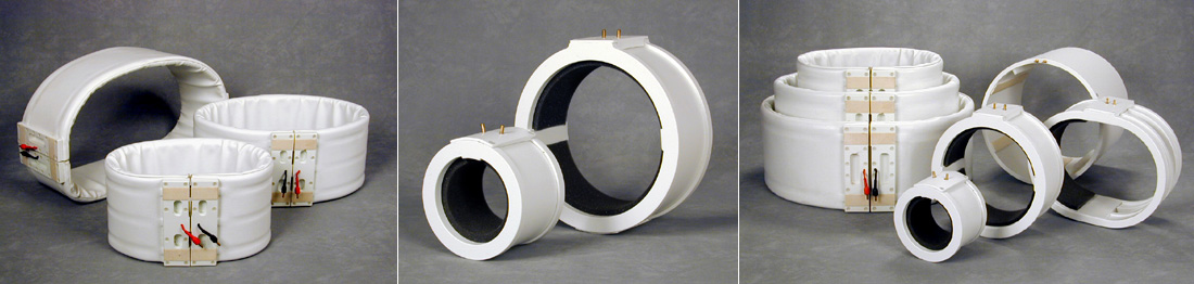
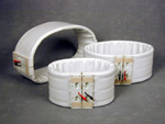
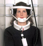 Cervical Solenoid Coil
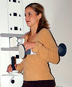
FONAR´S flexible Cervical Solenoid Coil optimizes imaging of the cervical spine. In the photo, the patient´s head was immobilized
for a cervical spine study in the neutral position. The coil is also ideally suited for Position Imaging™ (pMRI™) applications
of the cervical spine, including flexion, extension, rotation, and lateral bending.
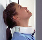 Expanded Diameter Solenoid Cervical Coil
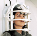
The Expanded Diameter Solenoid Cervical Coil is for patients that require
a more loosely fitting coil in order to flex, extend or bend their necks
as far as they are able to. pMRI™ applications give the referring
physician the most complete picture of the patient´s condition.
 High–performance Spine and Body Coil Set
(45–inch, 55–inch and 65–inch circumferences) These flexible wraparound coils provide uniform posterior–to–anterior signal intensity and
extended longitudinal coverage for spine and body imaging. The user can choose the optimal patient filling–factor to increase SNR.
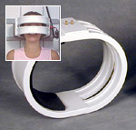 Signal Plus(TM) Open Head Coil
The Signal Plus™ Open Head Coil employs an advanced multi–conductor design that provides the high Signal–to–
Noise Ratios necessary for neuroimaging applications.
 Signal
Plus™ Knee
Signal
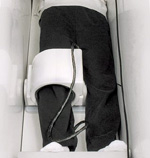
Plus™ Knee
This circular multi–conductor receiver coil for knee imaging can also
be used for the thigh, calf and ankle.
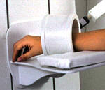 Wrist Coil
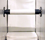
The FONAR wrist coil is a rigid, multi–conductor receiver coil optimized for high–resolution wrist or small extremity imaging.
PLANAR COILS
The unique transaxial horizontal magnetic field of the Upright™ MRI makes it the ONLY Open MRI system that can use flat planar coils. The advantage of planar coils is that they can be placed close to the targeted anatomy, which results in high Signal–to– Noise Ratios (SNR) and, therefore, excellent images.
 Planar T–L (Thoracic–Lumbar) Coil
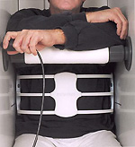
The Planar Thoracic–Lumbar Coil is placed directly behind the patient´s back. The patient
leans against the coil and sits comfortably throughout the scan.
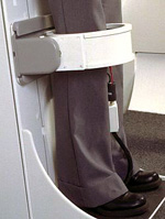
 Shoulder Coil – Seated
FONAR´s shoulder coil employs a unique double–planar design.
The coil, which can be used on either shoulder, interfaces with an immobilization
fixture that attaches to the patient bed.
QUADRATURE COILS
The quadrature coil combines two coils, called a quadrature pair, configured to operate in concert to achieve higher SNR than can be obtained with a single receiver coil.
 Quadrature Head Coil
The Quadrature Head Coil provides excellent neuroimaging capabilities. It
can be used with the patient in either an upright or recumbent posture.
Adjustable movement inhibitors keep the patient´s head immobilized
throughout the scan. The large spaces between the coil conductors allow
the patient an unobstructed view of a TV mounted on the scanner room wall.
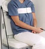
 Quadrature T–L (Thoracic-Lumbar) Coil
Because of its unique magnet configuration, the Upright™ MRI is the
ONLY MRI scanner that is able to utilize a solenoid (wrap-around) coil and
a flat planar coil as its quadrature pair. Each coil in FONAR´s quadrature
pair offers its own special advantage: The solenoid coil is remarkably SNR–efficient
— more so than saddle–shaped coils used with conventional superconductive
MRI scanners; and the companion planar coil achieves remarkable SNR because
of its close proximity to the targeted anatomy. The combination of the two
makes the Quadrature Thoracic–Lumbar Coil an extraordinary performer.
 VersaRest™ Fixture
The VersaRest™ fixture provides the patient with something sturdy
to lean on during the scan, making the patient comfortable and reducing
body motion. For example, for flexion studies of the lumbar spine, patients
bend forward at the hip and comfortably rest their forearms on the VersaRest™
fixture. The VersaRest™ fixture can be placed anywhere in the patient
gap, and is easily installed or removed in a few seconds.
 Quadrature Knee Coil
The Upright™ MRI is the ONLY MRI scanner capable of performing weight–bearing
extremity studies. Weight–bearing knee scans can be done with the
patient standing or with the bed tilted back to any selected angle. With
the bed tilted back, the patient is usually more comfortable and is better
able to remain still throughout the procedure. The coil attaches directly
to the bed. Patient positioning is quick and easy.