Studies Using the FONAR UPRIGHT® Multi-Position™ MRI
Published in The Journal of Neurology Demonstrates the Value of
FONAR UPRIGHT® Imaging in Evaluating the Extent of Hypermobility
in Chiari Malformation Patients
MELVILLE,
NEW YORK, March 13, 2008 - FONAR Corporation (NASDAQ-FONR),
The Inventor of MR Scanning, announced that the value of the FONAR
UPRIGHT© Multi-Position™ MRI in the diagnosis and
evaluation of Chiari malformation patients has just been published
by The Chiari Institute, North Shore-Long Island Jewish Health
Systems, in the Journal of Neurosurgery: Spine, December 2007,
Volume 7
http://thejns.org/doi/abs/10.3171/SPI-07/12/601 and www.northshorelij.com/workfiles/chiari/J%20Neurosurg%20Spine%20article%20Dec%2007.pdf
The
authors were Thomas H. Milhorat, M.D., Paolo A. Bolognese, M.D.,
Misao Nishikawa, M.D., of the Department of Neurosurgery, The
Chiari Institute, North Shore-Long Island Jewish Health Systems;
Nazli B. McDonnell, M.D., PhD., of the NIH National Institute
on Aging; and Clair A. Francomano, M.D. of the Greater Baltimore
Medical Center. The article, titled, “Syndrome of occipitoatlantoaxial
hypermobility, cranial settling, and Chiari malformation Type
I in patients with hereditary disorders of connective tissue”,
examined patients with CT as well as the FONAR UPRIGHT® MRI.
The
conclusion of the study was to report a previously unrecognized
association between Chiari Malformation Type I (CM-I) and Hereditary
Disorders of Connective Tissue (HDCT). The study occurred between
January 2002 and April 2007 and involved 2, 813 patients, of which
45% were referred for evaluation after failed Chiari Malformation
surgery.
 |
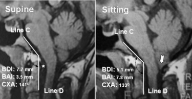
| FIG. 6. Results of vertical MR imaging
in a 27-year-old woman with HDCT/CM-I. Midsagittal image
in supine position (left) showing normal basion–dens
interval (7.7 mm), normal basion– atlas interval (3.5
mm), normal clivus–axis angle (141°), large retroodontoid
pannus, and low-lying cerebellar tonsils. On assumption
of the upright position (right), there is evidence of cranial
settling (2.6 mm decrease of basion–dens interval),
posterior gliding of occipital condyles (4.3 mm increase
of basion–atlas interval), anterior flexion of the
occipitoatlantal joint (8° decrease of clivus–axis
angle), increased basilar impression, and cerebellar ptosis
with downward displacement of cerebellar tonsils to C-1
(white arrow). Note the greatly increased impaction of the
foramen magnum anteriorly and posteriorly. Line C, superior
plane of the clivus; Line D, plane of the posterior surface
of the dens. Asterisk indicates the retroodontoid pannus. |
The
primary diagnostic tools utilized in the study were 2D reconstructed
CT and upright X-ray radiography. The final stage of the study
included examinations of patients in the FONAR UPRIGHT® MRI
for comparison.
The
authors described, for the first time, the phenomenon of “cranial
settling”, occurring in patients with both Chiari Malformation
1 (CM-I) and Hereditary Disorders of Connective Tissue (HDCT).
They
reported, “Recent experience with vertical MR imaging has
proved helpful in understanding the dynamic features of occipitoatlantoaxial
hypermobility. As shown in Fig. 6, functional cranial settling
was associated with notable displacements that included reduction
of the basion–dens interval, posterior gliding of the
occipital condyles, anterior flexion of the occipitoatlantal
joint, increased
basilar impression, and cerebellar ptosis with downward displacement
of the cerebellar tonsils. These displacements are consistent
with the often-pronounced symptoms and signs of lower brainstem
dysfunction experienced by patients with cranial settling on
assumption of the upright position.”
Concluding this peer-reviewed
paper, the acknowledgment by Dr. Milhorat, et al. kindly reported, “We
thank Dr. Raymond V. Damadian (Fonar Corporation) for providing
technical assistance and supervision of patients undergoing
vertical MR imaging.”
Dr.
Damadian, president and chairman of FONAR said, “We are
appreciative of Dr. Milhorat and his team for recognizing the
FONAR UPRIGHT® MRI’s power to visualize the full cranial
settling, cerebellar ptosis, cerebellar tonsil descent and foramen
magnum impaction that occurs in the Chiari Malformation-I/HDCT
patients so they can be optimally addressed surgically.”
About The Chiari Institute
The Chiari Institute
is the world's first comprehensive, multi-disciplinary center
for the management of patients suffering from Chiari
Malformation (CM), a rare structural condition that affects
the cerebellum; syringomyelia, a chronic disease of the spinal
cord; and related
disorders.
The Chiari Institute was founded in 2001 by Dr.
Thomas H. Milhorat,
chairman of the departments of neurosurgery at North Shore University
Hospital in Manhasset, N.Y., and Long Island Jewish Medical
Center in New Hyde Park, N.Y., and represents the fruition of
his decade-long effort to establish an institution dedicated
to the treatment of these often misdiagnosed conditions.
While at the State University of New York Health
Science Center in Brooklyn, Dr. Milhorat, et al. published a
landmark study in the journal Neurosurgery, 1999 May;44(5):1005-17.
The article is titled “Chiari I Malformation redefined:
clinical and radiographic findings for 364 symptomatic patients”.
What Dr. Milhorat and his team discovered, using MRI, was that
cerebral spinal fluid (CSF) flow was restricted or blocked around
the cerebellar tonsils for patients with Chiari Malformation.
They also found that Chiari is a condition where the crowding
is due to a small posterior fossa region, rather than a large
brain. For a reference visit: www.conquerchiari.org/subs%20only/Volume%202/Issue%202(11)/Milhorat%202(11).asp
For
more information on The Chiari Institute visit: www.northshorelij.com/body.cfm?ID=6407
#
The Inventor of MR Scanning™,
Full Range of Motion™, pMRI™, Dynamic™, Multi-Position™,
True Flow™, The Proof is in the Picture™, Spondylography™ and
Spondylometry™ are trademarks
and UPRIGHT® and STAND-UP® are registered trademarks
of FONAR Corporation.
This release may include
forward-looking statements from the company that may or may not
materialize. Additional information on factors that could potentially
affect the company's financial results may be found in the company's
filings with the Securities and Exchange Commission.
###

